
Page 92 - 2024-bfw-MyersAP4e-TE
P. 92
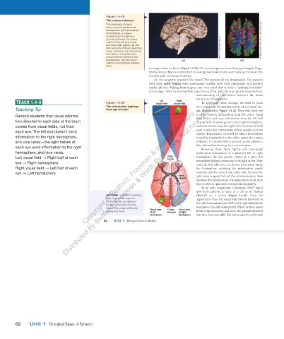
Figure   1.4-19
                                                           The corpus callosum
                                                         This large band of neural
                                                         fibers connects the two brain
                                                         hemispheres. (a) To photograph
                                                         this half-brain, a surgeon
                                                         separated the hemispheres
                                                         by cutting through the corpus                                           Dr. Patric Hagmann/CHUV, UNIL, Lausanne, Switzerland
                                                         callosum (see the blue arrow)
                                                         and lower brain regions. (b) This
                                                         high-resolution diffusion spectrum                   Martin M. Rotker/Science Source
                                                         image, showing a top-facing brain
                                                         from above, reveals the brain
                                                         neural networks within the two
                                                         hemispheres, and the corpus        (a)                        (b)
                                                         callosum neural bridge between
                                                         them.


                                                                             messages between them ( Figure 1.4-19 ). The neurosurgeons knew that psychologists Roger


                                                                             Sperry, Ronald Myers, and Michael Gazzaniga had divided cats’ and monkeys’ brains in this
                                                                             manner, with no serious ill effects.
                                                                                     So, the surgeons operated. The result? The seizures all but disappeared. The patients
                                                                             with these    split  brains   were surprisingly healthy, with their personality and intellect
                                                                             hardly affected. Waking from surgery, one even joked that he had a “splitting headache”
                                                                               ( Gazzaniga, 1967 ). By sharing their experiences, these patients have greatly expanded our
                                                                                                  understanding of interactions between the intact
                                                                                                  brain’s two hemispheres.
               TEACH 1.4-9                                         Figure   1.4-20    visual field  visual field        To appreciate these findings, we need to focus
                                                                                 Left
                                                                                         Right
                                                           The information highway                for a minute on the peculiar nature of our visual wir-
               Teaching Tip                              from eye to brain                        ing, illustrated in   Figure 1.4-20  Note that each eye

                                                                                                                     .


               Remind students that visual informa-                                               receives sensory information from the entire visual
                                                                                                  field. But in each eye, information from the left half
               tion directed to each side of the brain                                            of your field of vision goes to your right hemisphere,
               comes from visual fields, not from                                                 and information from the right half of your visual field
               each eye. The left eye doesn’t send                                                goes to your left hemisphere, which usually controls
                                                                                                  speech. Information received by either hemisphere
               information to the right hemisphere,                                               is quickly transmitted to the other across the corpus
               and vice versa—the right halves of                                                 callosum. In a person with a severed corpus callosum,
               each eye send information to the right                                             this information sharing does not take place.
                                                                                                           Knowing these facts, Sperry and Gazzaniga
               hemisphere, and vice versa.                                                        could send information to a patient’s left or right
                                                                                     Optic
               Left visual field   Right half of each                                nerves       hemisphere.  As the person stared at a spot, the
               eye   Right hemisphere                                                             researchers flashed a stimulus to its right or left. They
                                                                                                  could do this with you, too, but in your intact brain,
               Right visual field   Left half of each  Copyright © Bedford, Freeman & Worth Publishers.   the hemisphere receiving the information would
                                                                                                  instantly pass the news to the other side. Because the
               eye   Left hemisphere  Distributed by Bedford, Freeman & Worth Publishers. Not for redistribution.
                                                                                          Optic   split-brain surgery had cut the communication lines
                                                                                          chiasm
                                                                               Speech             between the hemispheres, the researchers could, with
                                                                                                  these patients, quiz each hemisphere separately.
                                                                                                       In an early experiment,  Gazzaniga (1967)  asked
                                                                                                  split-brain patients to  stare  at a dot as he flashed
                                                               split brain       a condition resulting   HE•ART on a screen (  Figure  1.4-21    ). Thus,  HE

                                                         from surgery that separates              appeared in their left visual field (which transmits to
                                                         the brain’s two hemispheres
                                                         by cutting the fibers (mainly            the right hemisphere) and ART in the right field (which
                                                         those of the corpus callosum)   Visual area  Corpus   Visual area  transmits to the left hemisphere). When he then asked
                                                         connecting them.    of left  callosum  of right  them to  say  what they had seen, the patients reported
                                                                             hemisphere   hemisphere  that they had seen ART. But when asked to  point  with
                                                        82   Unit 1  Biological Bases of Behavior
                                                  03_myersAPpsychology4e_28116_ch01_002_163.indd   82                               15/12/23   9:23 AM


               82   Unit 1  Biological Bases of Behavior


          03_HammerTE4e_47547_ch01_2a_163_4pp.indd   82                                                                         07/02/24   5:23 PM