
Page 75 - 2024-bfw-MyersAP4e-TE
P. 75
Module 1.4b
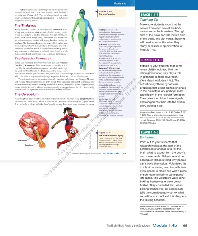
The brainstem is also a crossover point, where most nerves
to and from each side of the brain connect with the body’s FIGURE 1.4-7
opposite side (Figure 1.4-7). This peculiar cross- wiring — the The body’s wiring TEACH 1.4-5
brain’s contralateral hemispheric organization — is but one of
the brain’s many surprises. Teaching Tip
The Thalamus Make sure students know that the
nerves from each side of the body
Sitting atop the brainstem is the forebrain’s thalamus, a pair forebrain consists of the
of egg-shaped structures that act as the brain’s sensory control cerebral cortex, thalamus, and cross over in the brainstem. The right
center (see Figure 1.4-6). The thalamus receives information hypothalamus; manages complex side of the brain controls the left side
from all the senses except smell, and routes that information cognitive activities, sensory of the body, and vice versa. Students
to the brain regions that deal with seeing, hearing, tasting, and and associative functions, and
voluntary motor activities.
touching. The thalamus also receives some of the replies from brainstem the central core will need to know this when they
those regions, which it then directs to the medulla and to the of the brain, beginning where study hemisphere specialization in
hindbrain’s cerebellum. Think of the thalamus as being to sen- the spinal cord swells as it Module 1.4c.
sory information what Seoul is to South Korea’s trains: a hub enters the skull; the brainstem
through which traffic passes en route to various destinations. is responsible for automatic
survival functions.
The Reticular Formation medulla [muh-DUL-uh] the
hindbrain structure that is CONNECT 1.4-5
Distributed by Bedford, Freeman & Worth Publishers. Not for redistribution.
Inside the brainstem, between your ears, lies the reticular the brainstem’s base; controls
(“netlike”) formation. This nerve network, which is gov- heartbeat and breathing. Explain to your students that some
erned by the reticular activating system, extends from the spi- thalamus [THAL-uh-muss] research has indicated that the
nal cord right up through the thalamus. As the spinal cord’s the forebrain’s sensory control
sensory input flows up to the thalamus, some of it travels through the reticular formation, center, located on top of the reticular formation may play a role
brainstem; it directs messages to
Copyright © Bedford, Freeman & Worth Publishers.
which filters incoming stimuli and relays important information to other brain areas. the sensory receiving areas in the in dreaming (a topic covered in
The reticular formation also controls arousal — our state of alertness — as Giuseppe Moruzzi cortex and transmits replies to more detail in Module 1.5c). The
and Horace Magoun discovered in 1949. When they electrically stimulated a sleeping cat’s the cerebellum and medulla.
reticular formation, it almost instantly produced an awake, alert animal. When Magoun severed reticular formation a nerve activation-synthesis hypothesis
a cat’s reticular formation without damaging nearby sensory pathways, the effect was equally network that travels through the proposes that dream signals originate
dramatic: The cat lapsed into a coma from which it never awakened. brainstem into the thalamus;
it filters information and plays in the brainstem, and perhaps more
The Cerebellum an important role in controlling specifically, in the reticular formation.
arousal.
Extending from the rear of the brainstem is the hindbrain’s baseball-sized cerebellum; its cerebellum [sehr-uh- The cortex then takes those signals
name means “little brain,” which is what its two wrinkled halves resemble (Figure 1.4-8). BELL-um] the hindbrain’s and reorganizes them into the dream
The cerebellum (along with the basal ganglia — deep brain structures involved in motor “little brain” at the rear of the
brainstem; its functions include story as best it can.
processing sensory input,
coordinating movement output
and balance, and enabling Information from Hobson, J. A., & McCarley, R. W.
nonverbal learning and memory. (1975). Neuronal excitability amodulation over
the sleep cycle: A structural and mathematical
model. Science, 189(4196), 58–60. doi:10.1126/
science.1135627
Figure 1.4-8 TEACH 1.4-5
Tony Quinn/ZUMA Press/Newscom voluntary movements, as when Point out to your students that
The brain’s organ of agility
the cerebellum coordinates our
Cerebellum Hanging at the back of the brain, Enrichment
Spinal cord soccer player Mallory Pugh research indicates that part of the
controls the ball.
cerebellum’s function is to tell the
brain what to expect from the body’s
The Brain: Brain Regions and Structures Module 1.4b 65
own movements. Blakemore and her
colleagues (1998) studied why people
can’t tickle themselves. Volunteers lay
in a brain scanning machine with their
03_myersAPpsychology4e_28116_ch01_002_163.indd 65 15/12/23 9:23 AM
eyes closed. A plastic rod with a piece
of soft foam tickled the participants’
left palms. The volunteers were either
tickling themselves or were being
tickled. They concluded that, when
tickling themselves, the cerebellum
tells the somatosensory cortex what
sensation to expect and this dampens
the tickling sensation.
Information from Blakemore, S., Wolpert, D., &
Frith, C. (1998). Central cancellation of self-
produced tickle sensation. Nature Neuroscience, 1,
635–640.
The Brain: Brain Regions and Structures Module 1.4b 65
03_HammerTE4e_47547_ch01_2a_163_4pp.indd 65 07/02/24 5:22 PM