
Page 84 - 2024-bfw-MyersAP4e-TE
P. 84
Sensory Functions
If the motor cortex sends messages out to the body, where does the cortex receive incoming
messages? Penfield identified a cortical area — at the front of the parietal lobes, parallel to
and just behind the motor cortex — that specializes in receiving information from the skin
senses, such as touch and temperature, and from the movement of body parts. We now call
this area the somatosensory cortex. Stimulate a point on the top of this band of tissue and
a person may report being touched on the shoulder; stimulate some point on the side and
the person may feel something on the face.
The more sensitive the body region, the larger the somatosensory cortex area devoted
to it (see Figure 1.4-13). Your supersensitive lips project to a larger brain area than do your
toes, which is one reason we kiss rather than touch toes. Rats have a large area of the brain
devoted to their whisker sensations, and owls to their hearing sensations.
Scientists have identified additional areas where the cortex receives input from
senses other than touch. Any visual information you are receiving now is going to the
visual cortex in your occipital lobes, at the back of your brain (Figures 1.4-15 and 1.4-16).
If you have normal vision, you might see flashes of light or dashes of color if stimulated
in your occipital lobes. (In a sense, we do have eyes in the back of our head!) Having lost
much of his right occipital lobe in a tumor removal, a friend of mine [DM’s] was blind
to the left half of his field of vision. Visual information travels from the occipital lobes to
other areas that specialize in tasks such as identifying words, detecting emotions, and
Distributed by Bedford, Freeman & Worth Publishers. Not for redistribution.
recognizing faces.
Copyright © Bedford, Freeman & Worth Publishers.
Imperial College London (a) (b) Auditory Visual
cortex
cortex
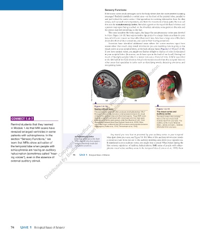
Figure 1.4-15
Seeing without eyes Figure 1.4-16
The psychoactive drug LSD often produces vivid hallucinations. Why? The visual cortex and
Because it dramatically increases communication between the visual auditory cortex
cortex (in the occipital lobe) and other brain regions. These fMRI scans The visual cortex in the occipital
CONNECT 1.4-7 show (a) a research participant with closed eyes who has been given lobes at the rear of your brain
a placebo and (b) the same person under the influence of LSD. Color receives input from your eyes. The
represents increased blood flow (Carhart-Harris et al., 2016). Other auditory cortex in your temporal
Remind students that they learned researchers have confirmed that LSD increases communication between lobes — above your ears — receives
brain regions (Preller et al., 2019; Timmermann et al., 2018). information from your ears.
in Module 1.4a that MRI scans have
revealed enlarged ventricles in some
patients with schizophrenia. In the Any sound you now hear is processed by your auditory cortex in your temporal
somatosensory cortex lobes (just above your ears; see Figure 1.4-16). Most of this auditory information travels
section “Sensory Functions,” we a cerebral cortex area at the front
of the parietal lobes that registers a circuitous route from one ear to the auditory receiving area above your opposite ear.
learn that MRIs show activation of and processes body touch and If stimulated in your auditory cortex, you might hear a sound. When taken during the
the temporal lobe when people with movement sensations. false sensory experience of auditory hallucinations, fMRI scans of people with schizo-
schizophrenia are having an auditory phrenia reveal active auditory areas in the temporal lobes (Lennox et al., 1999). Even
hallucination (sometimes called “hear- 74 Unit 1 Biological Bases of Behavior
ing voices”), even in the absence of
external auditory stimuli.
03_myersAPpsychology4e_28116_ch01_002_163.indd 74 15/12/23 9:23 AM
74 Unit 1 Biological Bases of Behavior
03_HammerTE4e_47547_ch01_2a_163_4pp.indd 74 07/02/24 5:22 PM