
Page 137 - 2024-bfw-MyersAP4e-TE
P. 137
Module 1.6b
2. Chemical reaction in turn
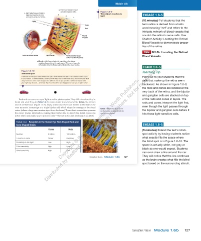
activates bipolar cells. Figure 1.6-9
1. Light entering eye triggers The retina’s reaction to
chemical reaction in rods 3 2 light ENGAGE 1.6-5
and cones at back of retina. 1
(10 minutes) Tell students that the
Light
term retina is derived from a Latin
Cone
Rod word meaning “net” and refers to the
Ganglion
Ganglion intricate network of blood vessels that
cell
cell
Bipolar
Bipolar
cell
cell Neural nourish the retina’s nerve cells. Use
impulse
Light Student Activity: Locating the Retinal
Light
3
Blood Vessels to demonstrate proper-
2 ties of the retina.
1 M1.6b: Locating the Retinal
Cross section of retina Optic nerve To the brain’s visual Blood Vessels
cortex via the thalamus
3. Bipolar cells then activate the ganglion cells, whose
combined axons form the optic nerve. This nerve transmits
Distributed by Bedford, Freeman & Worth Publishers. Not for redistribution.
information (via the thalamus) to the brain’s visual cortex.
TEACH 1.6-5
Figure 1.6-10 Teaching Tip
The blind spot
There are no receptor cells where the optic nerve leaves the eye. This creates a blind spot Point out to your students that the
Copyright © Bedford, Freeman & Worth Publishers.
in your vision. To demonstrate, close your left eye, look at the black dot, and move your face
away until one of the cars disappears. (Which one do you predict it will be?) Repeat with cells that make up the retina seem
your right eye closed — and note that now the other car disappears. Can you explain why?
backward. As shown in Figure 1.6-9,
the rods and cones are located at the
very back of the retina, and the bipolar
and ganglion cells are stacked on top
Rods and cones are our eyes’ light-sensitive photoreceptors. They differ in where they’re of the rods and cones in layers. The
found and what they do (Table 1.6-1). Cones cluster in and around the fovea, the retina’s rods and cones interpret the light first,
area of central focus (Figure 1.6-8). Many cones have their own hotline to the brain: One even though the light passes through
cone transmits its message to a single bipolar cell, which relays the message to the visual fovea the central focal point
cortex (where a large area receives input from the fovea). These direct connections preserve in the retina, around which the the bipolar and ganglion cells before it
the cones’ precise information, making them better able to detect fine detail. Cones can eye’s cones cluster. hits those light-sensitive cells.
detect white and enable you to perceive color — but not in the dark (Sabesan et al., 2016).
TABLE 1.6-1 Receptors in the Human Eye: Rod-Shaped Rods and
Cone-Shaped Cones ENGAGE 1.6-5
Cones Rods (5 minutes) Extend the text’s blind-
Number 6 million 120 million spot activity by having students notice
Location in retina Center Periphery what exactly fills the space where
the blind spot is in Figure 1.6-10. The
Sensitivity in dim light Low High
Color sensitivity High Low Omikron/Science Source space is actually white, not gray or
black as one would expect. Students
Detail sensitivity High Low can even draw a line around the car.
They will notice that the line continues
Sensation: Vision Module 1.6b 127
as the brain creates what fills the blind
spot based on the surrounding stimuli.
03_myersAPpsychology4e_28116_ch01_002_163.indd 127 15/12/23 9:25 AM
Sensation: Vision Module 1.6b 127
03_HammerTE4e_47547_ch01_2a_163_4pp.indd 127 07/02/24 5:27 PM